Integrating Clinical Science, Digital Technology, and Comprehensive Patient Assessment
Modern diagnosis requires more than examining individual teeth or treating isolated symptoms. The Institute’s approach integrates clinical examination, digital imaging, cephalometric analysis, TMJ evaluation, occlusal assessment, airway considerations, and functional relationships into a comprehensive diagnostic framework. The Institute combines advanced diagnostic technologies with proprietary software and structured clinical protocols to help clinicians develop a more complete understanding of the patient and establish a logical connection between diagnosis, treatment objectives, treatment mechanics, and documentation.
The Institute’s integrated approach presents a unified diagnostic technology platform and clinical methodology.
- JKL Software: Connecting the Diagnostic Data
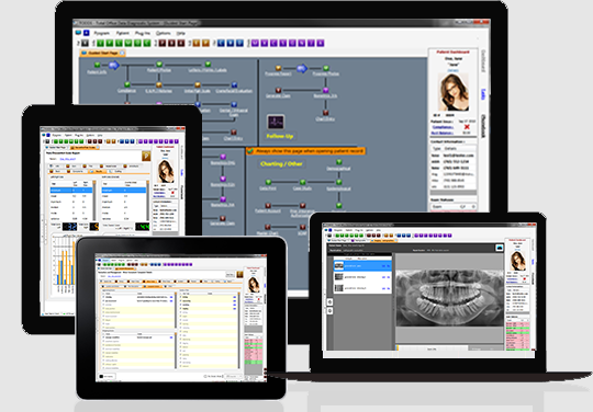
The JKL Software Program is designed to organize and analyze multiple clinical and diagnostic data points within a structured treatment-planning process.
Rather than evaluating each finding independently, the software helps clinicians identify relationships among:
- TM joints and condylar position
- Occlusion and mandibular position
- Cephalometric relationships
- Craniofacial structures
- Dental and skeletal relationships
- Airway considerations
- Functional and neuromuscular findings
- Clinical examination findings
- Treatment objectives and anticipated changes
The objective is to transform individual diagnostic findings into a coherent clinical picture that can support diagnosis and treatment planning.
From Data to Clinical Decisions
Clinical Data → Digital Analysis → Diagnostic Interpretation → Treatment Objectives → Treatment Plan → Documentation
This workflow is intended to improve the organization, consistency, and reproducibility of the diagnostic process while keeping the clinician—not the software—in control of clinical decision-making.
- Cephalometric Analysis
Cephalometric analysis provides a systematic method for evaluating skeletal, dental, and soft-tissue relationships.
The Institute incorporates cephalometric analysis into treatment planning to help clinicians evaluate:
- Maxillary and mandibular relationships
- Vertical facial relationships
- Sagittal relationships
- Incisor position and inclination
- Facial growth patterns
- Soft-tissue relationships
- Airway-related anatomical relationships
- Changes before, during, and after treatment
The goal is not simply to obtain measurements, but to understand what those measurements mean within the patient's overall functional and structural relationships.
- CBCT and TMJ Imaging
Three-dimensional imaging can provide anatomical information that may not be visible on conventional two-dimensional radiographs.
When clinically indicated, CBCT and TMJ imaging can assist in evaluating:
- Condylar morphology
- Glenoid fossa anatomy
- Osseous changes
- Joint-space relationships
- Condylar position
- Skeletal asymmetry
- Craniofacial anatomy
- Structures relevant to airway assessment
Imaging findings should be interpreted together with the patient's history, clinical examination, symptoms, functional findings, and other diagnostic information.
Imaging Is a Diagnostic Tool—Not the Diagnosis
The Institute emphasizes that imaging should answer specific clinical questions. The purpose is not to collect images simply because technology is available, but to use appropriate imaging to support clinical diagnosis and treatment decisions.
- Integrated TMJ, Occlusion, Orthodontic, and Airway Assessment
A central principle of the Institute is that the oral and craniofacial complex functions as an interconnected system.
The diagnostic process therefore considers relationships among:
TM Joints ↔ Mandible ↔ Occlusion ↔ Muscles ↔ Craniofacial Structures ↔ Tongue ↔ Airway
A change in one component may influence other components. Treatment planning should therefore consider these relationships rather than treating each structure as an isolated problem.
This integrated approach is particularly important when evaluating patients with complex combinations of TMD, malocclusion, craniofacial imbalance, functional disorders, and airway-related concerns.
- Digital Treatment-Planning Workflow
The Institute's technology is organized around a structured diagnostic workflow:
Step 1 — Patient History
Collect relevant medical, dental, functional, and symptom information.
Step 2 — Clinical Examination
Evaluate the TM joints, muscles, occlusion, mandibular function, craniofacial relationships, oral habits, and other clinically relevant findings.
Step 3 — Diagnostic Imaging
Select appropriate radiographic and three-dimensional imaging based on the clinical questions being investigated.
Step 4 — Digital Analysis
Enter and organize relevant diagnostic measurements and findings using the Institute's digital tools.
Step 5 — Relationship Analysis
Evaluate how the individual findings relate to one another and identify potential contributing factors.
Step 6 — Diagnosis
Develop a comprehensive diagnosis based on the total clinical and diagnostic picture.
Step 7 — Treatment Objectives
Define what the treatment is intended to accomplish and establish measurable treatment goals.
Step 8 — Treatment Planning
Develop the appropriate sequence and mechanics for treatment based on the diagnostic findings.
Step 9 — Documentation
Generate organized clinical documentation supporting the diagnosis, treatment rationale, objectives, and proposed treatment.
Step 10 — Follow-Up and Evaluation
Compare treatment progress with the original diagnostic findings and treatment objectives.
- Medical Necessity and Clinical Documentation
One of the important applications of the Institute's digital system is the organization of medical-necessity documentation.
The JKL Software Program is designed to assist clinicians in connecting:
Diagnostic Findings → Clinical Diagnosis → Functional Problems → Treatment Objectives → Recommended Treatment
This creates a documented clinical rationale for why a particular treatment may be indicated.
Where appropriate, the resulting documentation may support clinical communication, insurance submissions, patient records, and legal or regulatory documentation requirements.
The software is intended to assist documentation; it does not independently establish medical necessity, determine insurance coverage, or replace the clinician's professional judgment.
- Technology That Supports the Clinician
The Institute's philosophy is simple:
Technology should enhance clinical judgment—not replace it.
Digital tools can improve the organization and analysis of complex information, but the final diagnosis and treatment plan remain the responsibility of the qualified clinician.
The Institute therefore teaches clinicians not only how to use technology, but how to understand the clinical principles behind the data.
- The Institute's Integrated Digital Diagnostic Model
The Institute brings these technologies together into a single diagnostic philosophy:
Examine → Measure → Image → Analyze → Integrate → Diagnose → Plan → Document → Monitor
This model is designed to help clinicians move from fragmented diagnostic information toward a comprehensive, systematic, and technology-supported understanding of the patient.
The Institute Difference
The Institute does not teach technology as an isolated skill. It teaches clinicians how to combine:
Clinical Examination
- Advanced Imaging
- Cephalometric Analysis
- TMJ Evaluation
- Occlusal Analysis
- Airway Assessment
- Digital Data Analysis
- Treatment Planning
- Comprehensive Documentation
into an integrated diagnostic and treatment-planning system.
The result is a technology-enabled clinical process designed to make diagnosis more systematic, treatment planning more transparent, and clinical documentation more comprehensive.